Exploring the fragility of a piglet’s gut

Enteric diseases, including the development of diarrhoea, are an often seen problem in raising piglets, causing severe economic losses due to premature death, reduced piglet performance as well as accumulating costs from treatment as well as diagnostic measures. But why are piglets so highly susceptible to germs which don’t affect older animals and why is their course of disease often much more dramatic?
When it comes to diarrhoea in piglets besides infections, four other factor complexes have to be considered (Table 1). The most important fact is that piglets are born with an immature gut system. The maturation of the gut starts immediately after birth and is influenced by some perinatal factors. The first sign to start the maturation is the increase of arterial oxygen at birth followed by the influence of certain hormones (e.g. cortisol and growth factors). So events like premature delivery or prolonged parturition leading to neonatal hypoxia may already increase the risk for intestinal dysfunction and enteric diseases as the maturation may already be disturbed.

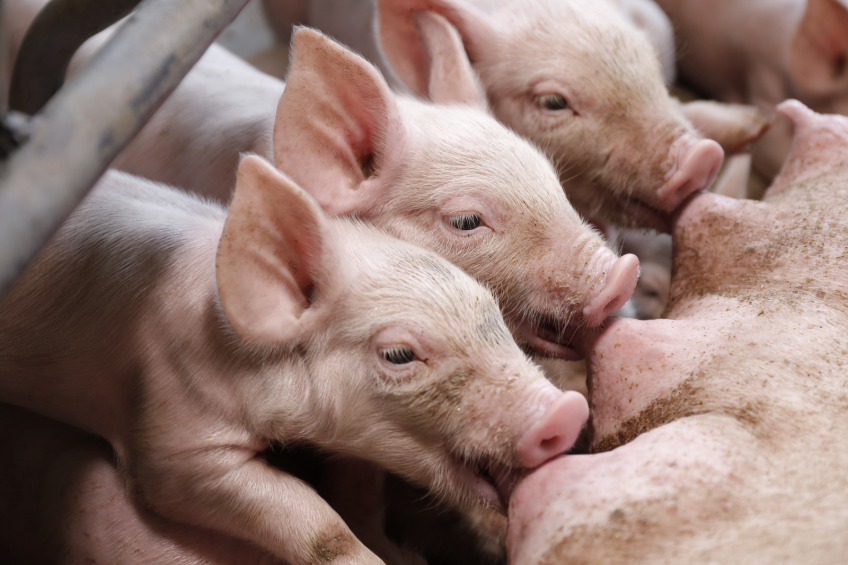
The next factor for the gut maturation is the presentation of enteral nutrients. This makes the susceptibility of the individual piglet towards intestinal diseases dependent on the amount, quality and composition of colostrum and milk intake as well as on the offering of solid feed components. Therefore the colostrum has a special function for the gastrointestinal condition of the piglet as it not only plays a role within the gut maturation but also provides immunoglobulins for the piglet’s immune system. These immunoglobulins are absorbed during the first 48 hours after birth for the maternal systemic immunity of the piglet or colonise the crypts (IgA) of the small intestine for a local immunity to control harmful intestinal infections during the development of the intestinal mucosal immune system. This development of the, at the beginning very rudimentary, local immunity takes place during the first 6 weeks of life and may be delayed by early weaning.
Physical and chemical changes during gut maturation
As the maturation of the gut proceeds there are some changes in physical and chemical status of the tissue between the newborn or weaned piglet and the adult pig. Firstly, the time needed to restore damaged gut tissue quickens with increasing age. The time for mucosal repair and replace lost or damaged villi and functioning epithelial cells is round about four to six days in piglets compared to two to three days in adult swine. Secondly, the permeability of the tight junctions is higher during the first hours of life. This fact is joined by a higher risk of absorption of dietary toxins or endotoxins. Linked to the different generations of enterocytes during maturation there are also differences in the produced digestive enzymes like HCl, amylases and lipases or lactase. Whereas some, like lactase, are highly produced in the newborn piglet and decline over the time, other enzymes like the mentioned amylases, lipases or HCl start on a very low level and increase substantially during the first 3-6 weeks of life. This means that the suckling and freshly weaned piglets are not able to use some diet components from feed for adult pigs if provided. These components may stay unused in the intestinal lumen providing nutrients for the growth of bacterial pathogens or leading to diarrhoea by an increased osmotic pressure and associated water within the intestinal lumen. And at least in accordance with the changing of the cell generations, the availability of receptors for pathogens expressed in the intestine change. Therefore some pathogens may only adhere to the epithelium of very young piglets or the piglets might become susceptible for specific pathogens over the time causing different susceptibilities and clinical pictures of disease at different ages of the pig. The most famous example is the receptor for fimbriae subtypes of Escherichia coli.
Pathomechanisms of diarrhoea in young pigs
Despite all these changes within the young pig at least all diarrhoeic diseases are based upon three different pathomechanisms: hypersecretion, malabsorption or inflammation. The malabsorptive diarrhoea or also osmotic form of diarrhoea is based upon the fact that nutrients cannot be absorbed and stay within the gut lumen. This may be due to the fact that they cannot be utilised as necessary because specific enzymes are missing, because there is an excessive supply or because mature cells capable of absorption are missing due to previous insults. The electrolytes and nutrient solutes retained in the lumen lead to an associated influx of water and therefore more liquid faeces.
The secretory form of diarrhoea is based upon a stimulation of secretion of electrolytes (mainly chloride) followed by the efflux of water within the crypt region of the small intestine due to infectious, neurological or toxic impacts. Inflammation leads to the development of diarrhoea by an increased oedema of the mucosa, increased mucosal permeability and mucosal necrosis. Additionally some inflammatory products also stimulate the secretion of electrolytes and H2O within a feedback mechanism.
Which are the most important infectious pathogens involved in piglet diarrhoea? In some cases diarrhoea is only an associated disease or secondary symptom and other pathogens might be of great importance for a particular husbandry condition e.g. the prevalence for Strongyloides ransomi decreased with the implementation of slatted floors.
Regarding all areas of piglet production and husbandry conditions the five most important infective pathogens for piglet diarrhoea remain Escherichia coli, Coronavirus infections, Clostridia ssp., Isospora suis and especially for weaned piglets Salmonella infection (see below‘The big 5 of piglet diarrhoea’). It should be mentioned that other diseases may be of special interest and importance other than the general conditions. Therefore, and because of the immature immunity of the piglet, it is most important to start further diagnostics as well as therapy as soon as diarrhoea occurs.
References available on request
The ‘big 5’ of piglet diarrhoea

Escherichia coli infection
Escherichia coli infections are important for piglets within the first days of life leading to the so called neonatal diarrhoea, or for weaning pigs causing post-weaning diarrhoea. Both clinical pictures are caused by enterotoxigenic Escherichia coli (ETEC). Enterotoxigenic Escherichia coli produce enterotoxins that induce secretory diarrhoea. They attach to the surface of the epithelium of the small intestine by fimbrial adhesins. These adhesins are the reason why there are the two clinical pictures of diarrhoea in piglets. As receptors for some fimbriae like F5 and F6 decreases during proceeding gut maturation, some receptors e.g. for F18 fimbriae are only expressed in older piglets starting at the age of two to three weeks. Produced toxins lead to electrolyte and water secretion that cannot be resorbed completely in the large intestine. Clinical signs in both cases are a watery, profuse, and whitish to yellow faeces. The colour may change towards some shades of brown or grey after weaning. Mortality in suckling piglets can reach up to 70% in affected litters by dehydration and emaciation resulting in deadly electrolyte imbalances but may only reach up to 25% in post-weaning cases. Some piglets are genetically resistant to F4 or F18 Escherichia coli infections due to a lack of the specific receptors.

Coronavirus infections
Coronavirus infections leading to piglet diarrhoea are the transmissible gastroenteritis virus (TGEV) and the porcine epidemic diarrhoea virus (PED). The transmission of both viruses occurs by faecal-oral-route and both infections lead to an enterocyte necrosis and severe villous atrophy within the small intestine. Marking is the thin and transparent gut wall during necropsy and the clinical sign of diarrhoea combined with vomiting piglets. As a consequence of the villous atrophy, malabsorption and maldigestion resulting in osmotic diarrhoea develops accompanied by severe and fatal dehydration. The clinical relevance of TGEV has decreased over the last years due, but PEDV and especially some highly virulent strains is counted among the re-emerging diseases with mortality rates up to >70% in piglets. Diarrhoea due to these strains may occur in all age groups.

Clostridia ssp. infections
Enteric clostridial infections are caused by Clostridium perfringens types A and C as well as Clostridium difficile but Clostridium perfringens types A and C are the most common causes of clostridial diseases in piglets. Sows or spores in the environment are most likely the source of infection. The infection with type C is characterised by haemorrhagic, necrotic enteritis with deep mucosal necrosis, severe inflammation and emphysema of the small intestine. In severe cases it might also extend to the caecum and proximal colon. Onset is most commonly at three days after birth but may also occur after the first 12 hours of life. The piglets show depression, bloody diarrhoea and abdominal pain. Acute cases may change to chronic cases with persisting diarrhoea for more than two weeks. Clostridium perfringens type A is a member of the normal porcine gut flora but can cause disease in neonatal as well as weaned piglets. Clostridium perfringens type A diarrhoea is not as fatal as type C diarrhoea but occurs more frequently. As Clostridium perfringens type A is not capable of producing the ß1- toxin, mucosal necrosis is not as deep as with type C infections and in some cases only secretory diarrhoea may occur. Typical clinical signs are non-haemorrhagic, mucoid diarrhoea and sometimes serositis.

Isospora suis
Isospora suis is a protozoan parasite and effects the jejunum and ileum and only in heavy cases parts of the caecum or colon. The disease is also called neonatal coccidiosis. The infectious stages infiltrate the enterocytes where they multiply. This leads to a necrotic enteritis of the epithelium layer combined with the loss of mature enterocytes which are replaced by a high number of immature cells. The ability of absorption by the mature cells is limited whereas the secretory activity of the immature cells is high. Disease develops within four to seven days after infection. The mortality is usually moderate but may be higher in cases of other co-infections.

Salmonella infection
The enteric salmonellosis, mainly caused by Salmonella typhimurium, usually affects pigs between the fifth and the 16th week of life. Lesions are predominantly found within the ileum, caecum and spiral colon. After attachment to epithelial receptors the bacteria are transported via vacuole to the basement membrane of the intestine leading to local inflammation and necrosis. Typical lesions are oedematous walls of the intestine, multifocal erosions covered with fibrinonecrotic debris and an enlargement of the mesenteric lymph nodes. The watery, yellow diarrhoea without mucus or blood is the result of the inflammation as well as an increased electrolyte efflux caused by produced endotoxins. Although the main focus for enteric salmonellosis is in older pigs and the course of the disease is usually mild to moderate, Salmonella infections are of special interest due to zoonotic potential and monitoring strategies at slaughter.


