Spondylitis is emerging in broilers

In World Poultry Vol. 23 No. 12, 2007, we described spondylitis caused by the bacterium Enterococcus cecorum as a newly emerging disease in broiler breeders in the US. Until recently the disease was only a problem in male broiler breeders, but now we are also seeing it in flocks of broiler chickens, with many birds in the flock described as having “leg problems”.
By Dr. Tahseen Aziz, Rollins Animal Disease Diagnostic Laboratory, Raleigh, NC, US, and Dr. H. John Barnes, College of Veterinary Medicine, North Carolina State University, Raleigh, NC, US.
In broiler flocks the problem begins between 30 and 40 days of age, often with a high morbidity rate resulting in culling of many birds in the flock. Almost all of the affected birds are males. Selective loss of males from the flock results in lowered average carcass weights at processing, and increased feed conversion rates.
Birds presented to the Animal Disease Diagnostic Laboratory showed clinical signs of leg paralysis. Typically, they were in ventral recumbency with their legs extended forward and spread laterally, and their feet slightly raised off the ground. Other birds were in ventral recumbency with their legs extended backward or severely spread to the sides.
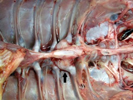
| Longitudinal section of the vertebral column of this 58-day-old broiler chicken shows the cavity in the body of deformed thoracicvertebrae (arrow) filled with necrotic debris and inflammatory exudate that compresses the spinal cord (SC). |
Necropsy examination of affected birds revealed a large, firm swelling in the ventral surface of the spine at the level of the articulating thoracic vertebra. Longitudinal section through the vertebral column revealed a cavity filled with necrotic debris in the body of the affected vertebrae. Due to the damage, the vertebra became deformed and compressed the spinal cord, and this was the cause of the leg weakness and paralysis.
| Here, 44- and 58-day-old broiler chickens show “leg weakness” due to spondylitis and subsequent spinal cord damage. The birds are in ventral recumbency with their legs extended, forward, backward, or laterally. In the top figure, the bird’s feet are slightly raised off the ground. | ||
| Here is a histologic section of the articulating thoracic vertebra of a 47-day-old broiler chicken. A space in the body of the articulating thoracic vertebra is filled with necrotic debrisand inflammatory exudate. The vertebra is deformed and there is marked compression of the spinal cord (SC) caused by the lesion. Numerous bacterial colonies are located within the lesion but they are not visible at this magnification. |


